Gallery
Overview
High Contrast and Resolution, Dual-Mode and Tunable Laser
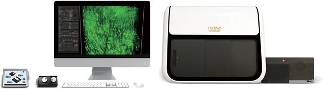
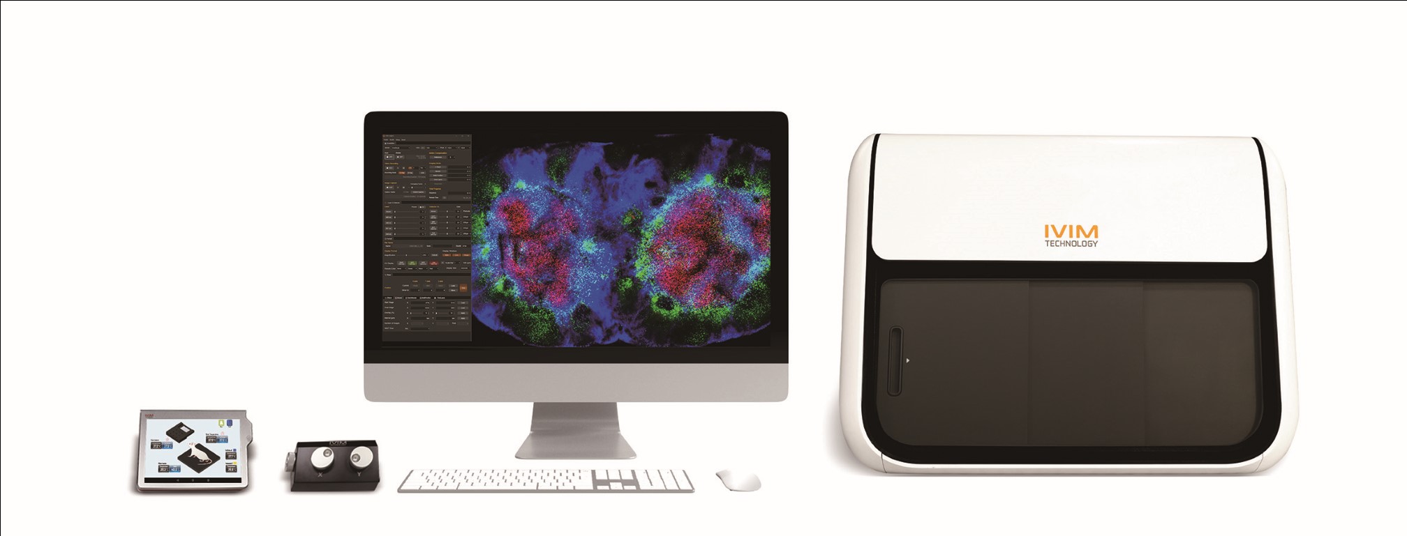
IVM-CM3 is a highly integrated All-in-One Intravital Microscopy system. It possesses the capability to focus on the desired wavelength using its tunable Two-Photon laser unit, covering wavelengths as low as 690 nm, reaching higher up to 1,050 nm, or anywhere in between. The IVM-CM3 seamlessly combines the advantages of both Confocal and Two-Photon Microscopy, offering endless possibilities for three-dimensional imaging of living cells near the skin or deep within tumors in small animals.
Key Features
■ Deep Tissue Imaging with a Tunable Long-Wavelength NIR fs-Laser System
■ One-Click Automated Transition between Confocal and Two-Photon Imaging Modes
■ Fully Integrated In Vivo Maintenance Unit / Animal Stage (e.g., Monitoring & Homeostatic Regulation of Animal Vitality)
■ Ultra High-Speed Imaging (max. 50 fps - 512 x 512 pixels)
■ 4D Animal Motion Compensation (XYZ & Time)
Specifications
|
Laser |
Confocal Laser Unit |
• 405 nm (20mW), 488 nm (20mW), 561 nm (20mW), 640 nm (20mW) |
|
Tunable Two-photon Laser Unit |
• Ti: Sapphire laser • Wavelength: 690 - 1050nm, Pulse width < 75 fs, Rep. rate: 80 MHz • Avg. power > 2.5 W, Dispersion compensation: 0 to -43,000 fs2 |
|
|
Fluorescence Detector |
Confocal Detector |
• Wavelength: 450 - 750 nm (DAPI, CFP, GFP, YFP, RFP, Cy5, Cy5.5, etc.) • 4 Ultra-broadband high SNR PMTs (UV to Near IR, Ultra High Sensitivity, Low Dark Current) • Single master pinhole |
|
Two-photon Detector |
• Wavelength: 450 - 750 nm (DAPI, CFP, GFP, YFP, RFP, Cy5, Cy5.5, etc.) • 4 high quantum efficiency PMTs (UV to Near IR, Ultra High Sensitivity, Low Dark Current) • Emission Filter: Individual filter can be mounted on each of four detectors |
|
|
Scan Head |
Scanner |
• Polygonal mirror (Fast axis scanning, Max. 66 kHz) • Galvano scanner (Slow axis scanning, Max. 200 ㎲/step) |
|
Imaging Head |
Objectives |
• Max. 5 objectives are mountable on IVM Engine Software controlled motorized turret (1X - 100X) • Compatible for commercial objectives |
|
Image |
FOV |
• 100 x 100 ㎛² - 10 x 10 mm² |
|
Pixel Resolution |
• Max. 2,048 x 2,048 pixels |
|
|
Imaging Speed |
• Standard: 30 fps @ 512 x 512 pixels • (Optional) High Speed: 50 fps @ 512 x 512 pixels |
|
|
Animal / Sample Stage |
Movable Stage |
• Travel Range: 50,000 x 50,000 x 75,000 ㎛ (XYZ) • Micromanipulation (Max. 0.2 ㎛ resolution) • 3-axis independent control with Jog Dial & IVM Engine software |
|
Specimen Holder |
• Flexible-design universal in vivo / ex vivo / in vitro specimen holders can be mounted • (Optional) Homeothermic warming system, Holders for window chamber |
|
|
Monitoring Camera |
• Real-time live animal / sample monitoring |
|
|
LED Light |
• Installed inside the machine to assist in the observation of live animals or samples |
|
|
Animal Motion Compensation (Tissue stabilization) |
4D In vivo Imaging Motion Compensation |
• XY motion compensation: Averaged image acquisition with motion artifact compensation • Z motion compensation: Image-based sample Z position adjustment for long-term intravital microscopic imaging & sample tracking (Feedback-loop automatic stage control) • T motion compensation: Image-based image XY position adjustment for long-term intravital microscopic imaging & sample tracking (Feedback-loop automatic stage control) • Combination of above three compensation for 4D in vivo motion compensation • Controllable by IVM Engine software |
|
Accessories Add-on |
Live Animal Maintenance Unit |
• Body Temp. Monitoring & Feedback Heater Control, including tablet PC • 4CH Rectal Probe, Body Plate Heater, Thermometer Sensor & Cover Glass Heater |
|
In vivo Imaging Chamber Sets |
• Dorsal Skinfold Chamber • Lung Imaging Chamber • Cranial Imaging Window • Abdominal Imaging Window • Pancreas Imaging Window • Mammary Imaging Window • Heart Imaging Chamber • Uterus Imaging Chamber |
|
|
Inhalation Anesthesia System |
• Whole Rodent Animal Inhalation Anesthesia System • Anesthesia Mask and Connections for Longitudinal Imaging |
|
|
Antibodies / Dyes |
• Fluorescent labeling agents, vascular dyes and conjugated antibodies |
|
|
Engine & Studio Software |
Image Display |
• Independent 4 single channel display (RGBA channel) • Overlay channel display (Selection among RGBA channel) |
|
In vivo Imaging Modes |
• Mosaic imaging (XY), Z-stack imaging (Z), Time-lapse imaging (T) • Time-lapse imaging at Multi-position (T - M) • Time-lapse & Z-stack imaging (TZ) • Time-lapse & Z-stack imaging at Multi-position (TZ - M) |